CE - what's that? - information for non-chemists
![]()
|
1. What is CE? | ||
| CE (capillary
electrophoresis) is quite a young analytical method having lost its
wallflower existence and nowadays is commonly accepted as a serious separation method.
Common principle of every electrophoretic separation is the phenomenon that under the
influence of an electrical field, electrically charged molecules (i.e. ions) tend to
wander towards the counterpolarized electrode (migration). The migration velocity usually
differs between different compounds and mainly depends on the size and the effective
charge of the analytes. Hence a mixture of samples can be separated into its constituents.
This principle is well established in bioanalytics for decades - but without capillaries -
where usually amino acids, peptides and proteins can thus be analysed. In a rather
"coarse" construction (at least from the analytical point of view!), a filter
paper is soaked with an electrically conductive salt solution (electrolyte) and the sample
is applied in a small band. When a high voltage is applied between two electrodes at the
ends of the paper, the sample molecules start to migrate - positively charged molecules
(cations) towards the negatively charged electrode (cathode), and negatively charged
molecules (anions) towards the positive electrode (anode). This flat-bed electrophoresis
shows some serious disadvantages: Most of all, the separation efficiency is very
bad, other points are the poor reproducibility (open system with a large surface),
difficult detection (mostly, the analytes have to be coloured with spray reagents), large
sample volumes and bad automaticability.
Abb. 1: separation principle It was in the Mid-80s when it was possible for the first time to transfer this separation principle from a slab gel into a capillary made from a special modification of silicon dioxide (fused-silica). It is now the same separation mechanism, but it takes place within this capillary that is filled with an electrolyte and whose ends are dipped into two buffer vials containing the electrodes, too. But in the special case of CE, another migration mechanism occurs additionally, the so-called electroosmotic flow (EOF). In a nutshell, it is the wall charge (zeta potential) of the silica that makes the buffer cations with their solvent shell tow the whole buffer liquid towards the cathode. This effect is only measurable in these small capillary dimensions since the charged capillary surface is very large compared to the electrolyte volume. Hence, in Free-Zone Capillary Electrophoresis (CZE), two different migration mechanisms overlap, with varying effects on the analytes. With a cathodic EOF, cations will be accelerated towards the cathode; however, anions will be slowed down by the EOF - fast anions will be moving slowlier to the anode, slow anions will even be tugged to the cathode despite their negative charge (fig. 1).
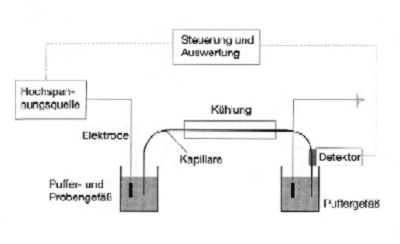
Abb. 2: schematic view of a separation device Fig. 2 shows the schematic principle of a CE device. Typical capillary dimensions are 30 - 70 cm in length and inner diameters of 25 - 100 µm. Most general applied voltages lie between 5 and 30 kilovolts. The sample is introduced at one end just by pressure or by a short voltage. The detection takes place at the opposite end, mostly done by measuring the absorption of UV light with a UV detector. The method impresses not only because of its huge separation power but also due to low detection limits, the small amount of sample necessary for an analysis and the high degree of automatisation. A special advantage is the reduced effort for sample preparation wgich is very useful in biochemistry. As uncharged compounds are transported only by the EOF, unwanted matrix substances can be separated from the target analytes mostly just by differences in their electrophoretic mobilities. Mankind is taking profit from this separation method in many ways. Just take a simple blood analysis: Whilst ca. 50 ml of blood received with intravenous cannulas were necessary for a medical check-up 15 years ago, you will need only a drop of blood from your finger tip today - and I guess it is no overstatement that the decoding of the human genome was only possible thanks to CE. |
|||
| More detailed information on CE (capillary electrophoresis) and CEC (capillary electrochromatography) are available at http://www.ceandcec.com | |||
... to be continued ...! |
|||
|
|||